MRI - Magnetic Resonance Imaging
|
Edit from "MRI Indication for the referring physician" |
||

|
Following is a quick overview of the main components of the MRI Hardware System. These include: |
||
|
THE MAIN MAGNET |
||
|
RECEIVER |
||
|
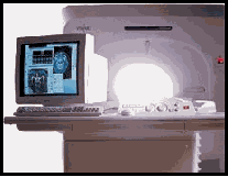
COMPUTER |
||
|
GRADIENT SYSTEM |
||

|
RF COILS |
||
|
|
||

|
TRANSMITTER |
||
|
|
||
|
Research Team |
||